Antibody Purification with Affinity Chromatography
by Simon Currie, Ph.D.

by Simon Currie, Ph.D.
Antibody purification leverages their affinity for immobilized Protein A, Protein G, or Protein L to isolate antibodies from complex mixtures. Proteins A, G, and L bind to different types of antibodies, so you should carefully consider which one will work best for the antibody that you’re purifying.
Antibodies are amazing! Antibodies from our immune cells help keep us healthy and mount a defense when we are infected. Scientists frequently use antibodies as research tools. And doctors even prescribe custom made antibodies to treat diseases including several types of cancers. Antibodies are purified for these scientific and medicinal applications.
In separate articles we have discussed affinity tags that are used to purify proteins such as his-tags and GST-tags. Antibodies, however, don’t need additional affinity tags added because they are already preloaded with a few different options for affinity purification!
Antibody purification leverages their affinity for immobilized Protein A, Protein G, or Protein L to isolate antibodies from complex mixtures. Proteins A, G, and L bind to different types of antibodies, so you should carefully consider which one will work best for the antibody that you’re purifying.
In this article we’ll first go over some general background about antibodies. Then we’ll use our antibody knowledge to cover how Protein A, Protein G, and Protein L purifications work. Lastly, we’ll discuss choosing the best protein for purifying your antibody.
General Features of an Antibody
Monoclonal vs Polyclonal Antibodies
Which Protein Should I Use to Purify My Antibody?
Before we get into antibody purification, we need to briefly describe:
Antibodies are incredibly complex. They consist of two bigger “heavy chain” proteins and two smaller “light chain” proteins. Disulfide bonds form between the two heavy chains, and between each heavy chain and a light chain to form the characteristic Y shape that antibodies are known for (Figure 1).
Each light chain consists of one variable domain and one constant domain, whereas each heavy chain is made up of one variable domain and three to four constant domains.
The variable domains of the heavy and light chains reside next to each other and form the surface of the antigen-binding fragment (Fab) that recognizes the molecular bits of foreign pathogens (Figure 1). An antigen is the molecule that the antibody binds to.
In contrast, the crystallizable fragment (Fc) consists of only constant domains. The hinge region connects the Fab and Fc fragments and is where the two heavy chain antibodies are bonded together.

Figure 1. Structural features of an antibody. Two heavy chains (purple) are disulfide bonded to each other (gray lines), and to two light chains (orange). The heavy and light chains make up the antigen binding fragment (Fab), whereas only the heavy chain is present in the crystallizable fragment (Fc). Two antigen binding sites (circled) are present on the tips of the Fab.
There are five classes or isotypes of antibodies in humans: IgA, IgD, IgE, IgG, and IgM. Ig is short for immunoglobulin, and antibodies are a type of immunoglobulin. The different classes of antibodies have distinct types of heavy chains. Heavy chains a, d, e, g, and m form IgA, IgD, IgE, IgG, and IgM classes, respectively.
Different antibody classes are found in different locations in our bodies, recognize distinct antigens, and have different biological functions. See Table 1 for a breakdown of what makes each of these classes unique.
Table 1. Antibody Classes
|
Class |
Heavy Chain |
Unique Features |
|
IgA |
a |
Prevents pathogen colonization in saliva, tears, breast milk, and on mucosal membranes. |
|
IgD |
d |
Found on naïve B cells that have not yet been exposed to antigens. |
|
IgE |
e |
Protect against parasitic worms, also involved in allergies and asthma. |
|
IgG |
g |
Detect pathogens in blood and extracellular fluid. |
|
IgM |
m |
Initiate inflammatory reactions to neutralize and clear pathogens. |
Monoclonal antibodies are generated by identical immune cells which are all originally derived from a single parent cell. This means that each monoclonal antibody is identical and recognizes the same binding site, or epitope, on the antigen of interest (Figure 2).

Figure 2. Monoclonal antibodies (purple and orange) are identical and bind to the same site on an antigen (blue). Polyclonal antibodies (slightly different shades of purple and orange) bind to the same antigen, but often via different sites.
Polyclonal antibodies come from a collection of immune cells in a host animal. This means that polyclonal antibodies are a group of distinct antibodies that all bind to the same antigen (Figure 2). However, different types of antibodies within that collection often bind to different epitopes on the antigen.
So which type of antibody is best? It depends on your intent.
Monoclonal antibodies are used when it’s important to have identical molecules that bind to a single antigenic epitope with high specificity. For example, therapeutic antibodies usually need to bind to an exact site on a protein to block its function; binding anywhere else likely will not provide the desired therapeutic benefit. For this reason, monoclonal antibodies are most often the better choice for antibody drug development.
Polyclonal antibodies are cheaper and faster to produce, and they are more tolerant to changes in the antigen such as epitope denaturation or slight variations in protein sequence. Since they recognize multiple epitopes, polyclonal antibodies are better for detecting scarce proteins. However, polyclonal antibodies are notoriously prone to batch-to-batch variability. Unfortunately, you never know exactly which collection of polyclonal antibodies you are working with!
Polyclonal antibodies also have higher rates of cross-reactivity, which is when they bind to a protein other than the desired antigen. See the last section of this article for an example of cross reactivity leading to false interpretations and years of lost research productivity!

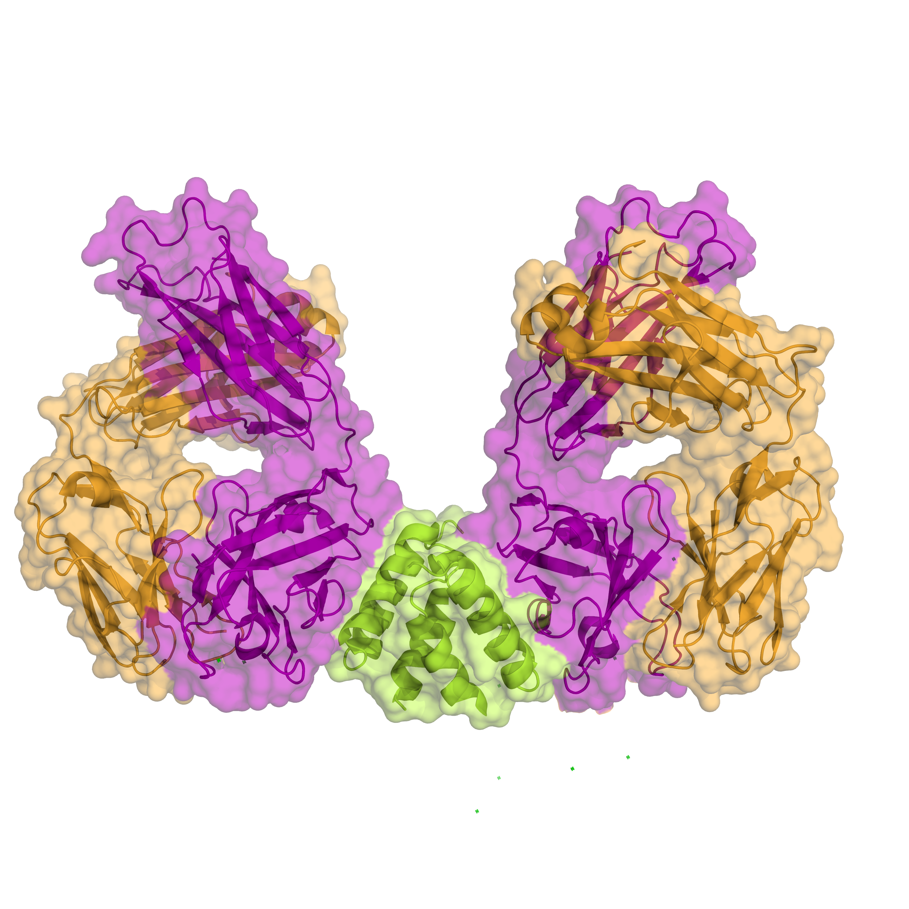
Figure 3. Protein A (green) bound to human IgM Fab via the heavy chains (purple). Light chains are also shown in orange (PDB: 1DEE).
For further details about monoclonal and polyclonal antibodies, and how they are produced, check out this article.
Phew! Now that we’ve covered the basics of antibodies, let’s jump into different options for affinity purifying them.
Protein A is a bacterial protein from the cell wall of Staphylococcus aureus that binds to the heavy chains of antibodies (Figure 3). Traditionally protein A was purified from S. aureus cultures. However recombinant protein A expressed in Escherichia coli is frequently used today.
Native or recombinant protein A is conjugated to agarose beads. Antibody-containing solutions are then loaded onto a protein A column. After washing, the antibodies are then eluted by adding buffer with acidic pH (pH<3) (Figure 4).

Figure 4. Purification of antibodies. Affinity-tagged proteins bind to agarose beads conjugated with interacting partner molecule such as protein A (column 2). After washing, antibodies are eluted with an acidic pH elution buffer that weakens the interaction between the antibody and protein A (column 3).
Protein A is particularly useful because a pH gradient can be used to separate different antibodies in a polyclonal mixture (Sheng and Kong, 2012). That is, instead of doing a one-step elution, the pH can be slowly changed in order to separate different antibodies from one another (Figure 5).
Antibodies have poor long-term stability in acidic conditions, so the eluted antibodies are then pH adjusted back to neutral values (~ pH 7 to 8).

Figure 5. One-step elution (left) vs gradient elution (right). Protein A is particularly useful for separating different antibody species using a gradient elution.
This same general purification scheme is also used for proteins G and L. So, in the next sections we will just cover the unique features of these proteins and how they purify antibodies.
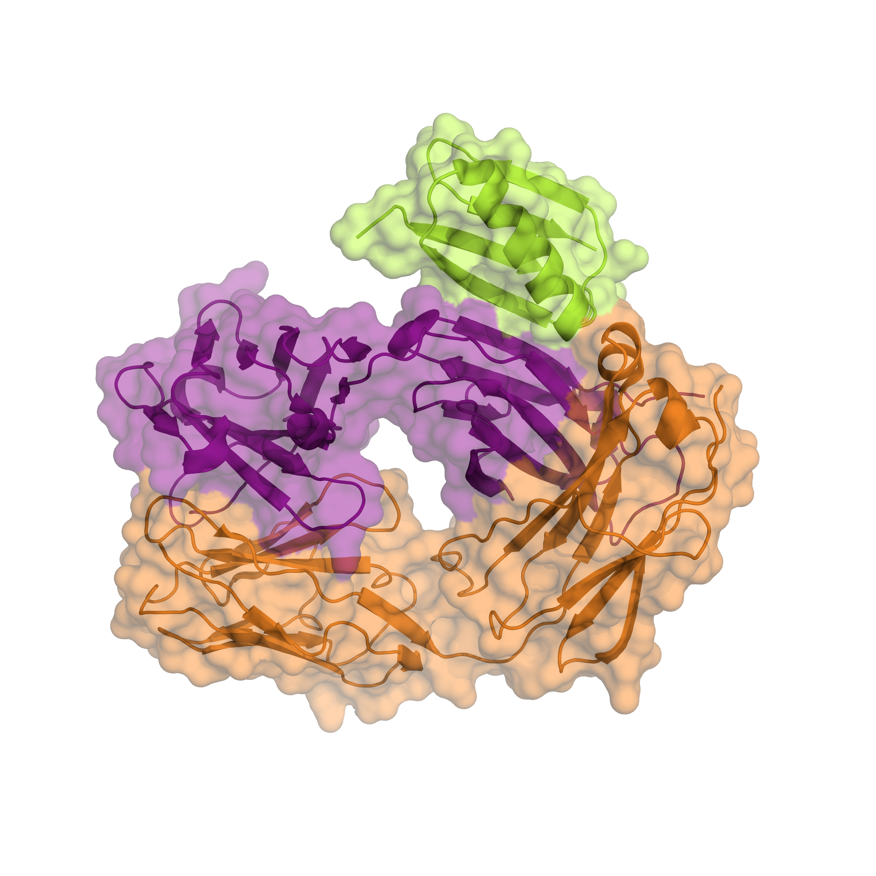
Protein G is a cell surface protein from Streptococcal bacteria that binds to antibodies (Figure 6). Like protein A, protein G also binds to the heavy chains of antibodies.
Native protein G also binds albumin with high affinity, making serum albumin a major contaminant of antibodies that are purified in this manner. However, recombinant protein G has been engineered to disrupt albumin binding, so there is no concern for albumin contamination when recombinant protein G is used.
The B domain of protein G is incredibly soluble. So much so that this domain is often used as a solubility tag to help keep other insoluble proteins in solution (Cheng and Patel, 2004).

Figure 6. Protein G (green) binding to IgG Fab light chain (orange) and heavy chain (purple) (PDB: 1IGC).
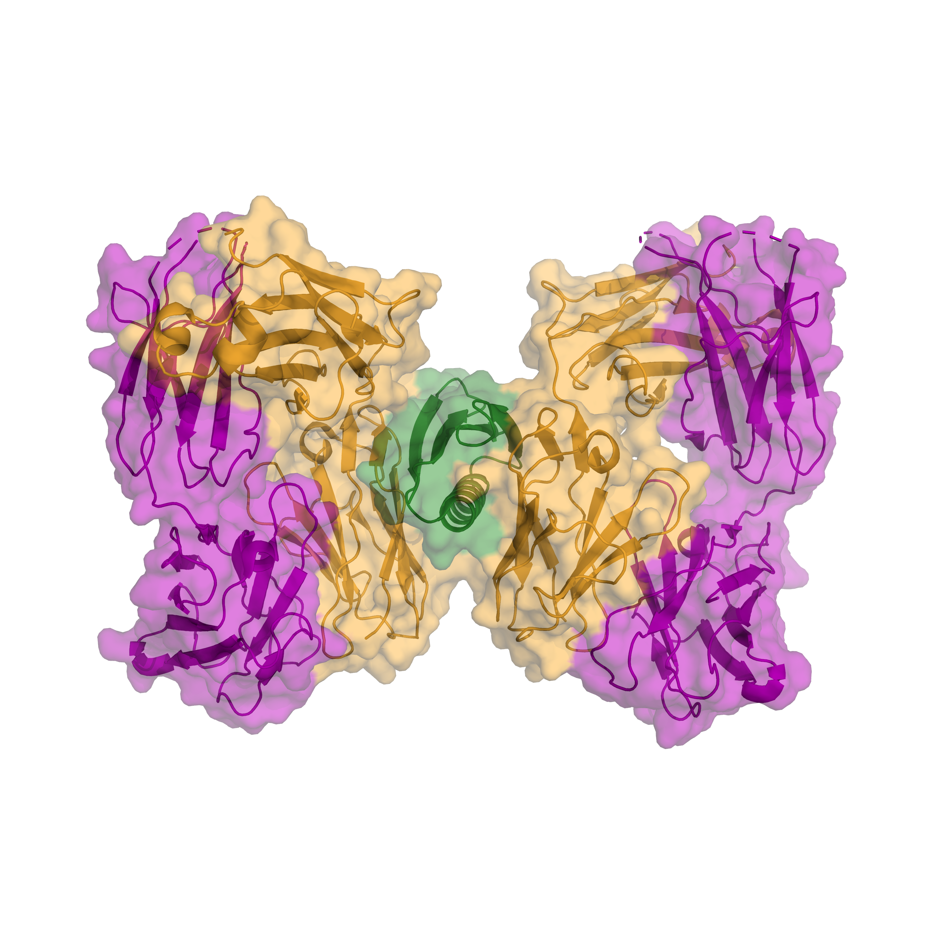
Protein L is a bacterial protein first purified from Peptostreptococcus magnus. Unlike proteins A and G, protein L binds to antibodies through the light chain (Figure 7). Protein L recognizes the kappa form of antibody light chains.
There is also a recombinant form of protein L, however, compared to proteins A and G this recombinant form has only existed for a fewyears (Kittler et al, 2022). Due to the newness of recombinant protein L, most commercially available protein L is the native form.
This different mode of engagement makes protein L more restrictive in terms of which species it will bind antibodies from. In the next section we’ll cover whichtypes of antibodies protein A, G, and L are preferred for.

Figure 7. Protein L (green) bound to human IgM Fab light chains (orange). Heavy chains are also shown in purple (PDB: 1HEZ).
Now that we’ve discussed the different proteins for affinity purifying your antibody, which one should you use?
For human and mouse IgG class antibodies, any of the above proteins should work well (See figure 8 below).
Protein L binds to all classes of human, mouse and rat antibodies, and it is the superior choice for IgA, IgD, and IgM classes in these species.
When it comes to IgG antibodies from other species, you’ll want to use either protein A or protein G. Protein A is the best choice for pig, dog, cat, and guinea pig IgGs. Conversely using protein G is ideal for goat, sheep, donkey, cow, and horse IgG antibodies. Both protein A and G work well for rabbit IgGs (Sheng & Kong, 2012).
That’s a lot to keep track of! In addition to Figure 8, this chart will help you decide which protein to use based on the type of antibody you are working with.
This article is intended to be an overview of antibody affinity purification, and as such we’ve only skimmed the surface here in terms of how to technically purify antibodies. For more details and step-by-step protocols, check out the following resources:

Figure 8. Antibody species and class recognition by proteins A, G, and L.

IPTG and auto-induction are two ways to induce protein expression in bacteria. They work similarly, but have different trade-offs in terms of convenience. While IPTG...

The final concentration of IPTG used for induction varies from 0.1 to 1.0 mM, with 0.5 or 1.0 mM most frequently used. For proteins with...

A His-tag is a stretch of 6-10 histidine amino acids in a row that is used for affinity purification, protein detection, and biochemical assays. His-tags...

Competent cells such as DH5a, DH10B, and BL21 will maintain their transformation efficiency for at least a year with proper storage. It is important to...
